![Takeru Eye Clinic | Takatori Shopping Street, Sawara-ku, Fukuoka City [Nishijin Station / Fujisaki Station]](https://takeru-eye.com/wp-content/uploads/2022/10/takeru_logo_for-WP-header.png)
Months to more than a decade after LASIK, clouding can develop in the cornea. Even if you feel after surgery that “there are no more problems,” the interface beneath the flap remains for life, and inflammation or fluid accumulation can occur there for some reason.
In this article I explain the types, diagnosis and treatment of late-onset corneal opacity.
The LASIK flap stays for life
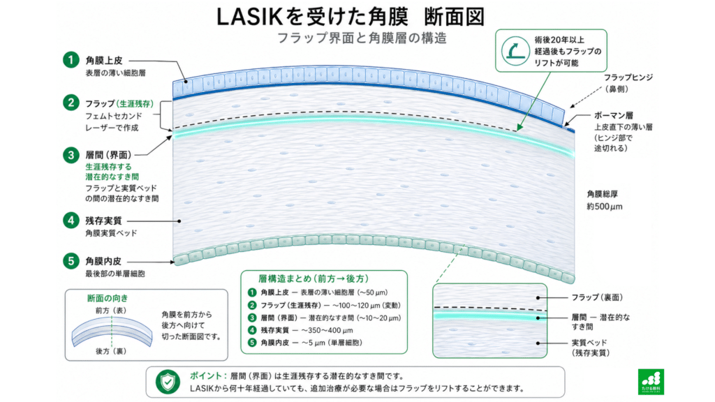
In LASIK, a thin “lid” (flap) is made on the surface of the cornea, and a laser is applied to the stroma (the main body of the cornea) beneath it to correct refraction. After surgery the flap is returned to its original position and adheres to the surface within a few hours.
However, the flap does not “heal completely and disappear.” An extremely thin “potential gap” between the flap and the underlying corneal stroma remains for decades after surgery¹.
In fact, it has been confirmed that the flap can be physically lifted even in corneas more than 20 years after LASIK¹. This may sound surprising, but it is a natural result given the structure of the surgery. Think of a cut made in a cake that does not disappear with time.
This gap normally causes no problems. But when some trigger is added, such as eye injury, eye inflammation, a change in eye pressure, ultraviolet light, or a change in general health, something abnormal can occur in the interface.
What is late-onset corneal opacity?
Late-onset corneal opacity after LASIK is not a single disease. It includes several conditions with different causes, and the treatment policy for each differs greatly. I will first show the overall picture and then look at each in detail.
The main conditions are these four: late recurrence of diffuse lamellar keratitis (DLK), pressure-induced stromal keratopathy (PISK), epithelial ingrowth, and stromal opacity (haze). Although they look similar, the treatments can be opposite, so accurate differentiation is extremely important.
Late recurrence of diffuse lamellar keratitis (DLK)
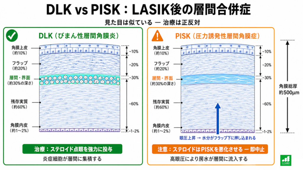
Diffuse lamellar keratitis (DLK) is a condition in which inflammatory cells gather beneath the LASIK flap. Because a white opacity like scattered sand is seen in the interface, it is also called “Sands of the Sahara syndrome.”
It is widely known as something that occurs early after surgery (days to weeks), but recurrence several years, or even more than ten years, after surgery has also been reported².
Why does it recur after so many years? When the surface of the cornea (the epithelium) is injured, cytokines (proteins that convey information between cells) that cause inflammation are released. The released cytokines draw inflammatory cells into the interface through the gap beneath the flap, the “path of least resistance”².
It is like a “passage” made more than 10 years ago continuing to serve as a shortcut for inflammatory cells.
Reported triggers of late-onset DLK include external force to the eye from sports, viral conjunctivitis, epithelial injury from severe dry eye, and changes in immunity accompanying systemic disease². In recent years, infection-related immune dysregulation of the ocular surface was reported to have triggered DLK in a case 20 years after surgery, again showing that the interface can permanently function as a site of inflammation².
Pressure-induced stromal keratopathy (PISK)
Pressure-induced stromal keratitis (PISK) is a condition in which a rise in eye pressure pushes water from the inside of the cornea into the gap beneath the flap³.
Although it looks very similar to DLK, the treatment is the opposite. For DLK, steroid eye drops are used to suppress inflammation, but in PISK the cause is often a rise in eye pressure from steroid eye drops, and increasing the steroid in that case raises eye pressure further and worsens the condition³.
There is another important characteristic. Because of the flap and changes in the interface, a cornea after LASIK differs from the “typical cornea” assumed by the Goldmann applanation tonometer³¹⁴. For that reason, pressure may be measured lower than the real value, and in PISK this becomes a “diagnostic pitfall.”
Whenever steroid eye drops are used in an eye after LASIK, this property of eye pressure must be kept in mind.
How eye pressure comes out low in eyes after LASIK is explained in detail in another article.
Epithelial ingrowth
Epithelial ingrowth is a condition in which epithelial cells covering the surface of the cornea grow into the interface from the edge of the flap and proliferate.
It often occurs early after surgery, but it can also progress late, triggered by trauma several years after surgery or by flap lifting for retreatment⁴.
When the epithelial cells spread toward the center of the cornea, they cause decreased vision and irregular astigmatism. In rare cases, as it progresses, enzymes produced by the epithelial cells gradually dissolve the flap itself, a condition called keratolysis⁴.
The degree of progression is classified by grade. Cases with a well-defined border within 2 mm of the flap edge are observed, and those progressing toward the center or accompanied by flap melting need early treatment⁴.
Stromal opacity (corneal haze)
の発生メカニズム-筋線維芽細胞経路-corneal-haze-formation-myofibroblast-pathway-1024x576.png)
Stromal opacity after LASIK is less frequent than after PRK (photorefractive keratectomy), but it can occur in those who had correction of high myopia or who had problems with the flap⁵.
The transparency of the cornea is maintained by orderly collagen fibers and quiescent keratocytes. When the epithelial basement membrane is damaged and its repair is delayed, cytokines such as TGF-β (transforming growth factor-β) flow continuously into the stroma. This changes keratocytes into myofibroblasts, which produce an opaque extracellular matrix, appearing clinically as “opacity”⁵.
Ultraviolet light (UV-B) has been shown to strongly promote this fibrosis process⁶, which is one reason wearing sunglasses after surgery is recommended.
How to tell whether it is “cloudy”
:LASIKフラップ界面の可視化-as-oct-lasik-flap-interface-visualization-1024x576.png)
From here the content is somewhat technical, concerning examination and diagnosis in the eye clinic. It is enough to read it as a reference for your visit.
For an accurate diagnosis of late-onset corneal opacity, imaging tests play an important role in addition to observation with a slit lamp.
Anterior segment optical coherence tomography (AS-OCT) is a test that can non-invasively observe the inside of the flap in cross-section. In PISK, a “gap” filled with fluid in the interface can be seen, and in epithelial ingrowth, cell masses with high reflectivity are observed³. It is an important tool for objectively visualizing conditions that are hard to judge with a slit lamp alone.
In vivo confocal microscopy (IVCM) allows observation at the cellular level. In DLK many inflammatory cells are seen, whereas in PISK cellular components are scarce and fine water gaps are observed within the stroma³.
Also, densitometry using a Scheimpflug camera can express corneal transparency numerically as grayscale units (GSU). It can supplement subjective slit-lamp evaluation with an objective index and is also useful for tracking the effect of treatment.
By combining these tests, conditions that look alike but differ can be told apart, leading to appropriate treatment.
How is it treated?
Treatment differs greatly depending on the cause.
For DLK, strong steroid eye drops are the first choice. In severe cases, the flap is lifted and the interface is irrigated to remove inflammatory cells². In some cases, systemic steroids are also considered.
For PISK, the basic approach is to stop the steroid and lower eye pressure. Eye-pressure-lowering drugs such as beta-blockers and alpha-2 agonists are used. Once the eye pressure improves, the water accumulated in the interface is naturally absorbed over days to weeks by the pumping function of the corneal endothelium³.
For epithelial ingrowth, treatment is considered according to the effect on vision and the degree of progression. Surgery is performed in which the flap is lifted again and epithelial cells are carefully removed from both the stromal bed and the underside of the flap. To prevent recurrence, mitomycin C (MMC) may also be used, but the recurrence rate is reported to range widely from 0 to 36%, so careful follow-up is needed⁴.
For fixed stromal opacity, phototherapeutic keratectomy (PTK) using an excimer laser is one option. To prevent recurrence, MMC is applied during surgery⁷. MMC is often used at a concentration of 0.02%; a high concentration (0.04%) is more potent but raises the risk of toxicity to the corneal endothelium, so it is not usually recommended⁸.
What to watch for in daily life
Protection from ultraviolet light is desirable not only early after surgery but continued over the long term. A correlation between UV-B exposure and the development of corneal opacity has been shown⁶, and wearing sunglasses with UV protection outdoors after surgery is recommended.
Those using steroid eye drops must have their eye pressure checked over time. Because eyes with a history of LASIK tend to be measured with low eye pressure, careful observation that does not miss even slight changes is important³.
Also, no matter how many years have passed since LASIK, it is important to see an eye doctor promptly if any of the following symptoms appear.
- Sudden decline in vision
- Glare, or a red eye
- Vision that looks white and hazy, as if seen through sand
These can be signs of late-onset inflammation or infection. It may be necessary to let go of the feeling that “it’s been a long time, so I’m fine.”
Long-term follow-up after LASIK

The generation that had LASIK in the 1990s and 2000s is reaching the age when cataract surgery is needed. In such patients, the structural changes of the cornea from LASIK affect every aspect of cataract surgery¹.
In cataract surgery, an eye with a history of LASIK requires a different method from the usual for calculating intraocular lens (IOL) power. Also, the steroid eye drops used after cataract surgery can trigger a recurrence of PISK, so postoperative eye pressure management is especially important¹.
Flaps made with the microkeratomes of that time are known to be thicker and more likely to have irregular edges than those made with today’s femtosecond lasers. It is also known that in this older LASIK, symptoms such as dry eye, halos and glare tend to remain relatively often¹.
An eye that has had LASIK keeps its structural characteristics even decades later.
References
- Sahay P, Bafna RK, Reddy JC, Vajpayee RB, Sharma N. Complications of laser-assisted in situ keratomileusis. Indian J Ophthalmol. 2021;69(7):1658-1669. doi:10.4103/ijo.IJO_1872_20 https://pmc.ncbi.nlm.nih.gov/articles/PMC8374806/
- Moshirfar M, Durnford KM, Lewis AL, et al. Five-Year Incidence, Management, and Visual Outcomes of Diffuse Lamellar Keratitis after Femtosecond-Assisted LASIK. J Clin Med. 2021;10(14):3067. Published 2021 Jul 11. doi:10.3390/jcm10143067 https://pmc.ncbi.nlm.nih.gov/articles/PMC8304683/
- Cabral-Macias J, García-De la Rosa G, Rodríguez-Matilde DF, et al. Pressure-induced stromal keratopathy after laser in situ keratomileusis: Acute and late-onset presentations. J Cataract Refract Surg. 2018;44(10):1284-1290. doi:10.1016/j.jcrs.2018.06.053 https://pubmed.ncbi.nlm.nih.gov/30107965/
- Ting DSJ, Srinivasan S, Danjoux JP. Epithelial ingrowth following laser in situ keratomileusis (LASIK): prevalence, risk factors, management and visual outcomes. BMJ Open Ophthalmol. 2018;3(1):e000133. Published 2018 Mar 29. doi:10.1136/bmjophth-2017-000133 https://pmc.ncbi.nlm.nih.gov/articles/PMC5895975/
- Wilson SE. Corneal myofibroblasts and fibrosis. Exp Eye Res. 2020;201:108272. doi:10.1016/j.exer.2020.108272 https://pubmed.ncbi.nlm.nih.gov/33010289/
- Stojanovic A, Nitter TA. Correlation between ultraviolet radiation level and the incidence of late-onset corneal haze after photorefractive keratectomy. J Cataract Refract Surg. 2001;27(3):404-410. doi:10.1016/s0886-3350(00)00742-2 https://pubmed.ncbi.nlm.nih.gov/11255052/
- Nassaralla BA, McLeod SD, Nassaralla JJ Jr. Prophylactic mitomycin C to inhibit corneal haze after photorefractive keratectomy for residual myopia following radial keratotomy. J Refract Surg. 2007;23(3):226-232. doi:10.3928/1081-597X-20070301-04 https://pubmed.ncbi.nlm.nih.gov/17385287/
- Netto MV, Mohan RR, Ambrósio R Jr, Hutcheon AE, Zieske JD, Wilson SE. Wound healing in the cornea: a review of refractive surgery complications and new prospects for therapy. Cornea. 2005;24(5):509-522. doi:10.1097/01.ico.0000151544.23360.17 https://pubmed.ncbi.nlm.nih.gov/15968154/
- Hamilton DR, Manche EE, Rich LF, Maloney RK. Steroid-induced glaucoma after laser in situ keratomileusis associated with interface fluid. Ophthalmology. 2002;109(4):659-665. doi:10.1016/s0161-6420(01)01023-5 https://pubmed.ncbi.nlm.nih.gov/11927421/
- Morales P, Camos-Carreras A, Spencer F, Torras J, Diaz P. Late-onset diffuse lamellar keratitis 15 years after laser in situ keratomileusis. Can J Ophthalmol. 2017;52(3):e91-e93. doi:10.1016/j.jcjo.2016.11.010 https://pubmed.ncbi.nlm.nih.gov/28576227/
- Amano R, Ohno K, Shimizu K, Suzuki M, Aizawa D, Komatsu M. Delayed-onset diffuse lamellar keratitis.
Journal of the Japanese Ophthalmological Society 107 (4): 202-207, 2003 https://www.nichigan.or.jp/Portals/0/JJOS_PDF/107_202.pdf - Japanese Ophthalmological Society. Guidelines for refractive surgery (8th edition). Japanese Ophthalmological Society.
https://www.nichigan.or.jp/Portals/0/resources/member/guideline/refractivesurgery_8.pdf - 101st Annual Meeting of the Japanese Ophthalmological Society, Commissioned Report I: Ocular cell biology: actions of the transforming growth factor-beta (TGF-β) superfamily in ocular tissues. Journal of the Japanese Ophthalmological Society. 1997;101:927.
https://www.nichigan.or.jp/Portals/0/JJOS_PDF/101_927.pdf - Fournier AV, Podtetenev M, Lemire J, et al. Intraocular pressure change measured by Goldmann tonometry after laser in situ keratomileusis. J Cataract Refract Surg. 1998;24(7):905-910. doi:10.1016/s0886-3350(98)80041-2 https://pubmed.ncbi.nlm.nih.gov/9682108/