![Takeru Eye Clinic | Takatori Shopping Street, Sawara-ku, Fukuoka City [Nishijin Station / Fujisaki Station]](https://takeru-eye.com/wp-content/uploads/2022/10/takeru_logo_for-WP-header.png)

Worldwide, ICL surgery has been increasing, and we are seeing more patients who need examination of the eye after ICL surgery.
What to watch for in an eye with an ICL:
Long-term complications involve
- glaucoma
- cataract
LASIK— just like eyes that are candidates for it,

eyes that are candidates for ICL surgery
= myopic eyes
= eyes at high risk of glaucoma and cataract1
.
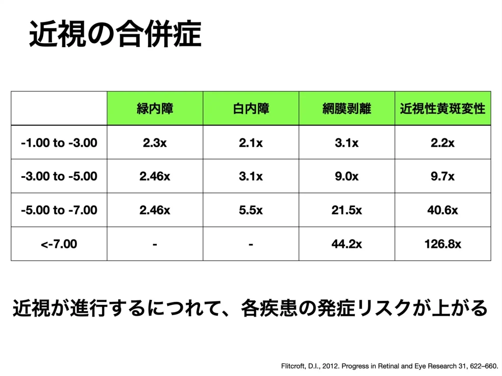
• Glaucoma
• Cataract
• Retinal detachment, etc.
ICL surgery is performed on eyes that already carry a high risk of eye disease in the future.
Long-term complications after ICL surgery (glaucoma, cataract, etc.) and their causes

Where the ICL lens is placed
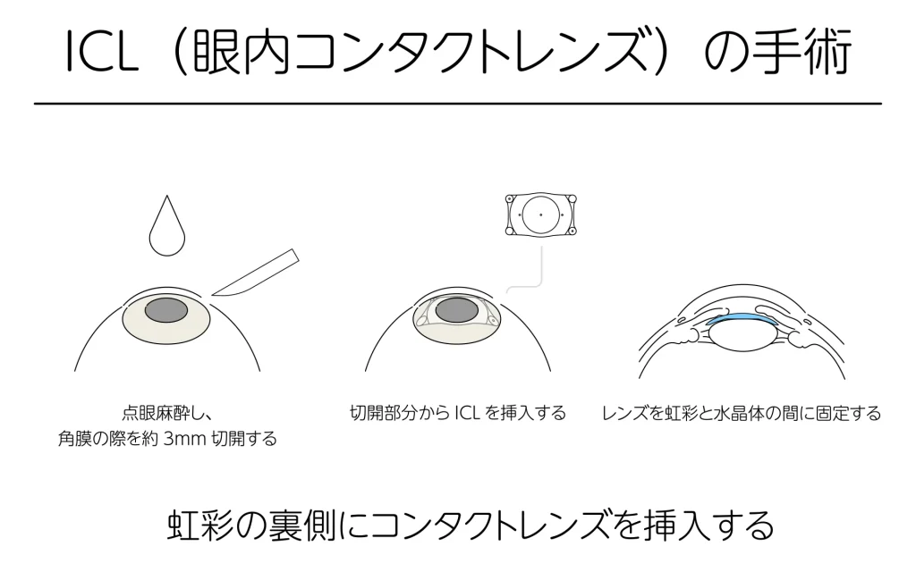
The ICL is inserted into the narrow space between the crystalline lens and the iris.
To give “good vision,” presbyopia (age-related loss of near focusing) sets in earlier.
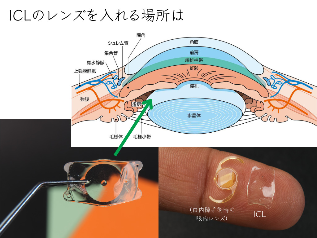
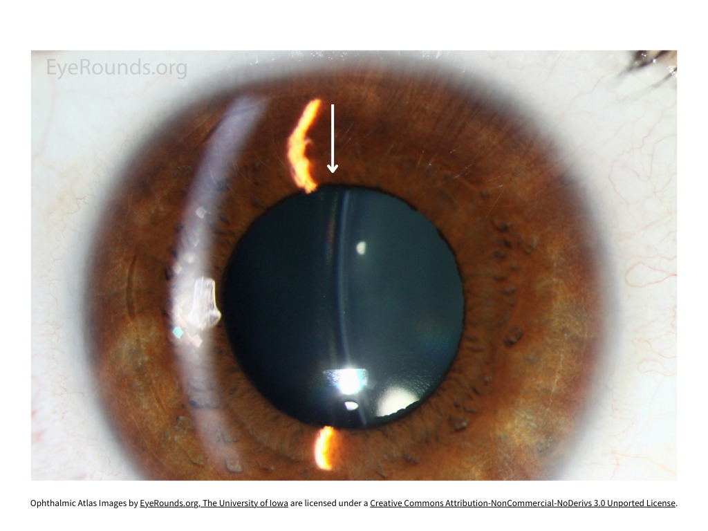
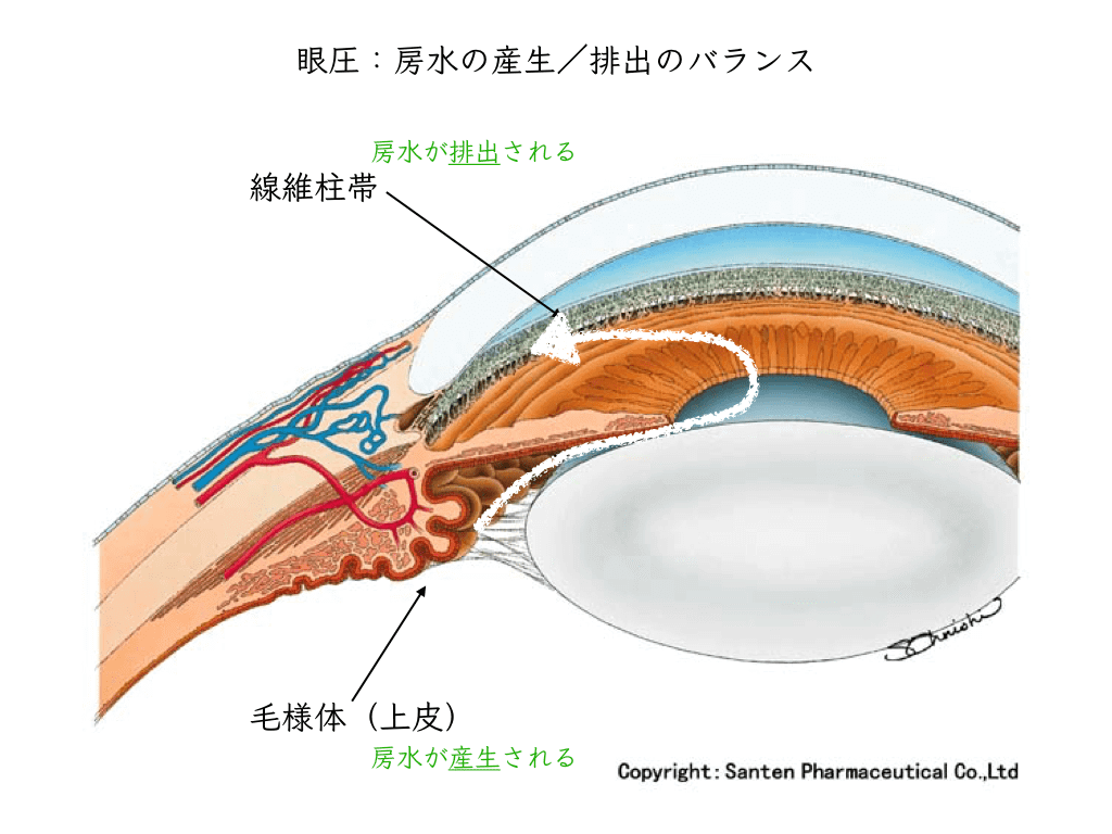
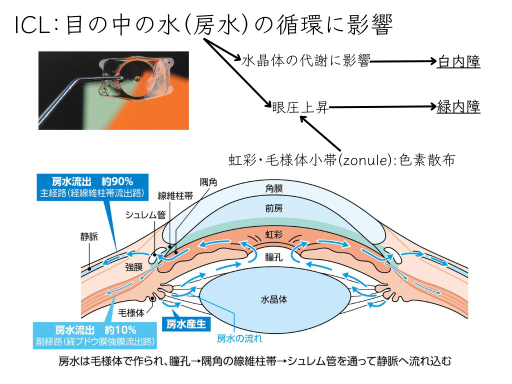
The ICL lens touches the iris and the zonule, and pigment is released and floats around inside the eye.
It clogs the angle, the drainage pathway for fluid in the eye, and eye pressure rises without you noticing.
Even when eye pressure rises, there are usually no symptoms.

As a result, changes in the visual field (narrowing and missing areas) gradually progress.
It proceeds without being noticed.
You notice the poor vision only when it is quite advanced
: the eye is by then in a state of advanced glaucoma2, 3。
This is glaucoma that has developed as a long-term aftereffect of the ICL.
Once glaucoma has reached an advanced stage, it cannot be reversed.
Also,
cataract after ICL surgery is thought to result from effects on aqueous humor circulation and, in turn, on lens metabolism3。
Unlike glaucoma, cataract can be treated with surgery to restore vision.
But the burden of cataract surgery, the instability of intraocular lens power calculation formulas, and difficulty with focusing remain as problems.
When cataract surgery is performed, the risks and complications of removing (explanting) the ICL are also an issue.
Risks and complications when removing (explanting) the ICL
After ICL surgery: if “something” happens after a long period,
Can the ICL be removed easily? What are the risks?
The incision is enlarged and the ICL is removed.
Video of ICL removal (translated summary)
- The ICL (Implantable Collamer Lens) is used in people with high myopia or myopic astigmatism, and is increasingly popular with growing demand for cosmetic results and refractive surgery.
- Before starting ICL removal, dilating the pupil to the maximum (maximal mydriasis) is important so that the haptics (the supporting arms of the lens) in the posterior chamber can be manipulated easily.
- A viscoelastic substance (especially a cohesive/dispersive one containing sodium hyaluronate and chondroitin sulfate) is injected into the anterior chamber. It helps maintain the anterior chamber properly.
- ICL removal is the process of engaging a hook in the dialing hole and gently pushing the four edges of the ICL into the anterior chamber (dislocation), taking care not to damage the corneal endothelium or the lens.
- Depending on the size of the corneal incision, the whole ICL is removed if the incision is 2.8 mm or larger, whereas if it is smaller than 2.8 mm, the ICL is cut in two before removal.
Various complications can occur when the ICL is pulled out (removed) from the eye4。
- (Traumatic) cataract
- Retinal tear / retinal detachment
- Glaucoma
- Corneal endothelial cellsloss
- Bullous keratopathy, etc.
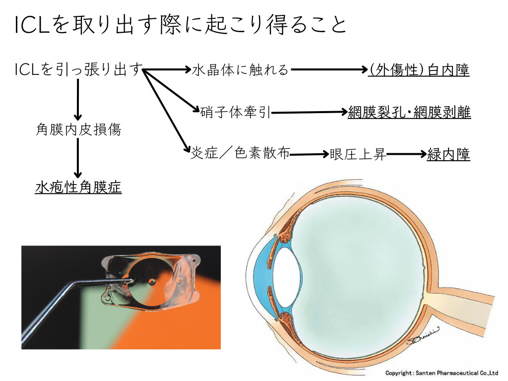
(Retinal tear: a condition in which “a hole opens in the retina”)
The most common complications when removing (explanting) the ICL are the development of cataract and an increased long-term risk of glaucoma.
In a 10-year study conducted in Switzerland, about 55% of patients developed cataract after ICL surgery, and 13% had ocular hypertension (raised eye pressure) requiring eye drop treatment5。


References
- Flitcroft, D.I., 2012. The complex interactions of retinal, optical and environmental factors in myopia aetiology. Progress in Retinal and Eye Research 31, 622–660. https://doi.org/10.1016/j.preteyeres.2012.06.004
- Sidhu, T., Desai, A., Gagrani, M., Dada, T., 2019. Phacomorphic glaucoma in a high myope with phakic intraocular lens. BMJ Case Rep 12, e227096. https://doi.org/10.1136/bcr-2018-227096
- Moya, T., Javaloy, J., Montés-Micó, R., Beltrán, J., Muñoz, G., Montalbán, R., 2015. Implantable Collamer Lens for Myopia: Assessment 12 Years After Implantation. J Refract Surg 31, 548–556. https://doi.org/10.3928/1081597X-20150727-05
- Zhang, H., Gong, R., Zhang, X., Deng, Y., 2022. Analysis of perioperative problems related to intraocular Implantable Collamer Lens (ICL) implantation. Int Ophthalmol 42, 3625–3641. https://doi.org/10.1007/s10792-022-02355-w
- Clinical Outcomes and Cataract Formation Rates in Eyes 10 Years After Posterior Phakic Lens Implantation for Myopia | Cataract and Other Lens Disorders | JAMA Ophthalmology | JAMA Network [WWW Document], n.d. URL https://jamanetwork.com/journals/jamaophthalmology/fullarticle/2498860