![Takeru Eye Clinic | Takatori Shopping Street, Sawara-ku, Fukuoka City [Nishijin Station / Fujisaki Station]](https://takeru-eye.com/wp-content/uploads/2022/10/takeru_logo_for-WP-header.png)

Floaters can sometimes accompany inflammation inside the eye (within the eyeball).
This article is about how posterior uveitis is discovered from the symptom of floaters.
Floaters That Reveal Inflammation Inside the Eye: Posterior Uveitis

Non-Inflammatory “Physiologic Floaters”
The Cause of Physiologic Floaters: “Posterior Vitreous Detachment”
Most floaters are caused by “posterior vitreous detachment.”
What causes the cloudiness inside the eye?
The membrane that surrounds the jelly-like vitreous, and the vitreous fibers themselves, are perceived as cloudiness.
In this case, there is no inflammation.
Inflammatory Floaters
Why Does Uveitis Cause Floaters?
When inflammation occurs inside the eye, inflammatory cells gather in the vitreous.
Clusters of immune cells (white blood cells) form and cause floaters.
Uveitis accompanied by cloudiness in the vitreous is called posterior uveitis.
Other causes of cloudiness inside the eye (vitreous) include
• Red blood cells from bleeding
• Retinal pigment epithelial cells that were located beneath a retina with a hole
There are two types of inflammation:
• Infectious (inflammation caused by microorganisms)
• Non-infectious (autoimmune: inflammation directed against one’s own tissues)
Is It Infectious Uveitis? “SSTTEEVE”
Is It Uveitis Caused by Microorganisms (“Infectious Uveitis”)?
Most infectious uveitis affects only one eye.
Infectious uveitis is sometimes discovered from floaters in just one eye.
I examine the inside of the eye with SSTTEEVE in mind1.
SSTTEEVE is an acronym for the following diseases:
- Sarcoidosis2
- Syphilis3
- Toxoplasmosis4
- Tuberculosis5
- Endogenous Endophthalmitis6
- Virus
: herpes simplex virus, varicella zoster virus (herpes zoster virus), cytomegalovirus - Et cetera
Based on the characteristic findings of each disease, decisions are made about the next tests and treatment.
PCR can confirm the presence of microorganisms7, 8.
Among other causes,
posterior uveitis caused by fungi (mold) or Toxocara also occurs, though rarely.
In addition, there is alsouveitis caused by anticancer drugs.
I have also seen a case in which the “proliferative membrane” of untreated, considerably advanced diabetic retinopathy was the cause of floaters.
Uveitis caused by tattoos9 also seems to exist, but I have never seen it.
Non-Infectious / Autoimmune Uveitis
Uveitis often reflects inflammation throughout the body.
This is why it is called the “internal medicine” of ophthalmology.
When the inflammation affects both eyes, non-infectious/autoimmune uveitis is considered.
○ Intestinal inflammation:
ulcerative colitis, Crohn’s disease
○ Spinal inflammation:
ankylosing spondylitis
○ Joint inflammation:
rheumatoid arthritis, juvenile idiopathic arthritis
○ Skin and mucous membrane inflammation:
psoriasis, Behçet’s disease
○ Multisystem inflammation:
sarcoidosis (lungs, lymph nodes, heart, eyes, etc.)
Behçet’s disease (mouth ulcers, skin inflammation, etc.)
After a medical interview, if necessary, care is provided in cooperation with an internist.
After a Hole Forms in the Retina (Retinal Tear / Retinal Detachment), Inflammatory Reactions Begin Inside the Eye
Retinal pigment epithelial cells are scattered from beneath the retina where the tear has formed.
Along with the tear, small blood vessels may rupture, resulting in vitreous hemorrhage.
» Floaters: the probability (risk) of retinal tear and retinal detachment

Other causes of floaters:
• Retinal pigment epithelial cells that were located beneath a retina with a hole
• Red blood cells from bleeding
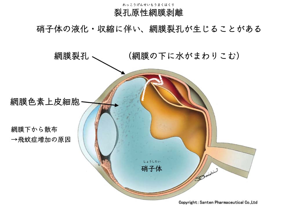
Retinal pigment epithelial cells produce large amounts of inflammatory proteins (cytokines).
A study at the Department of Ophthalmology, Kyushu University analyzed the vitreous of eyes with retinal detachment.
It was found to be inflammatory10.
It was also found that when the condition progresses to intractable proliferative vitreoretinopathy, inflammatory cytokines rise even further.
Proliferative vitreoretinopathy:
a condition in which proliferative membranes form after retinal detachment surgery, in retinal detachment due to a giant tear, or after retinal detachment is left untreated,
making it extremely difficult to treat.
It is thought to be caused by inflammatory changes driven by retinal pigment epithelial cells11.
Floaters caused by retinal tears or retinal detachment may also be inflammatory floaters.
(Retinal tears and retinal detachment are distinguished from posterior uveitis.)
Action is needed to prevent inflammation from progressing to proliferative vitreoretinopathy.
Prompt attention to the symptom of floaters is desirable.
Please Don’t Worry Too Much
Doctors make their judgment while imagining all of the above, including diagnosis by exclusion12.
Searching for medical information on your own and worrying about everything is excessive.
Floaters are very common in everyday ophthalmic practice.
Is a serious disease hidden?
Is it a sign of a bad disease?
An ophthalmologist can tell the risk by examining the inside of the eye with a microscope.
Examination without dilating the pupils can now reveal much of the inside of the eye.
Posterior uveitis sometimes requires not only eye drops but also injections around the eye and oral medication. The stages of treatment and side effects are explained in the next article.

References
- Mandelcorn, E.D., 2013. Infectious causes of posterior uveitis. Canadian Journal of Ophthalmology 48, 31–39. https://doi.org/10.1016/j.jcjo.2012.11.013
- Classification Criteria for Sarcoidosis-Associated Uveitis, 2021. . American Journal of Ophthalmology 228, 220–230. https://doi.org/10.1016/j.ajo.2021.03.047
- Oliver, G.F., Stathis, R.M., Furtado, J.M., Arantes, T.E., McCluskey, P.J., Matthews, J.M., Group, I.O.S.S., Smith, J.R., 2019. Current ophthalmology practice patterns for syphilitic uveitis. British Journal of Ophthalmology 103, 1645–1649. https://doi.org/10.1136/bjophthalmol-2018-313207
- Garweg, J.G., de Groot-Mijnes, J.D.F., Montoya, J.G., 2011. Diagnostic Approach to Ocular Toxoplasmosis. Ocular Immunology and Inflammation 19, 255–261. https://doi.org/10.3109/09273948.2011.595872
- Bansal, R., Basu, S., Gupta, A., Rao, N., Invernizzi, A., Kramer, M., 2017. Imaging in tuberculosis-associated uveitis. Indian J Ophthalmol 65, 264–270. https://doi.org/10.4103/ijo.IJO_464_16
- Sadiq, M.A., Hassan, M., Agarwal, A., Sarwar, S., Toufeeq, S., Soliman, M.K., Hanout, M., Sepah, Y.J., Do, D.V., Nguyen, Q.D., 2015. Endogenous endophthalmitis: diagnosis, management, and prognosis. Journal of Ophthalmic Inflammation and Infection 5, 32. https://doi.org/10.1186/s12348-015-0063-y
- Fallon, J., Narayan, S., Lin, J., Sassoon, J., Llop, S., 2022. The impact of polymerase chain reaction (PCR) on diagnosis and management of infectious uveitis at a tertiary care facility. Journal of Ophthalmic Inflammation and Infection 12, 1. https://doi.org/10.1186/s12348-021-00276-w
- Rothova, A., de Boer, J.H., ten Dam-van Loon, N.H., Postma, G., de Visser, L., Zuurveen, S.J., Schuller, M., Weersink, A.J.L., van Loon, A.M., de Groot-Mijnes, J.D.F., 2008. Usefulness of Aqueous Humor Analysis for the Diagnosis of Posterior Uveitis. Ophthalmology 115, 306–311. https://doi.org/10.1016/j.ophtha.2007.05.014
- Peterson, A.S., Patterson, A.W., 2022. Case Series: Tattoo-associated Uveitis. Optom Vis Sci. https://doi.org/10.1097/opx.0000000000001859
- Yoshimura, T., Sonoda, K.-H., Sugahara, M., Mochizuki, Y., Enaida, H., Oshima, Y., Ueno, A., Hata, Y., Yoshida, H., Ishibashi, T., 2009. Comprehensive Analysis of Inflammatory Immune Mediators in Vitreoretinal Diseases. PLoS ONE 4, e8158. https://doi.org/10.1371/journal.pone.0008158
- Dai, Y., Dai, C., Sun, T., 2020. Inflammatory mediators of proliferative vitreoretinopathy: hypothesis and review. Int Ophthalmol 40, 1587–1601. https://doi.org/10.1007/s10792-020-01325-4
- Trusko, B., Thorne, J., Jabs, D., Belfort, R., Dick, A., Gangaputra, S., Nussenblatt, R., Okada, A., Rosenbaum, J., for The Standardization of Uveitis Nomenclature (SUN) Project, 2013. The Standardization of Uveitis Nomenclature (SUN) Project: Development of a Clinical Evidence Base Utilizing Informatics Tools and Techniques. Methods Inf Med 52, 259–265. https://doi.org/10.3414/ME12-01-0063