![Takeru Eye Clinic | Takatori Shopping Street, Sawara-ku, Fukuoka City [Nishijin Station / Fujisaki Station]](https://takeru-eye.com/wp-content/uploads/2022/10/takeru_logo_for-WP-header.png)

“I was born prematurely and I’m told I had treatment for my eyes. They look normal, so I assume they’re fine—but do I still need to see an eye doctor now that I’m an adult?”
We hear this question at our clinic. In many cases, the answer is yes.
Retinopathy of prematurity (ROP) is often thought of as a condition of infancy. But whether someone was treated, or had mild disease that needed only observation, changes in the retina or vision can appear in adulthood.
Care does not end with the newborn period. Follow-up that continues from then on is the key to protecting vision in the future.
We would like to share the same message with parents who are worried about the eyes of a baby born prematurely.
Why we recommend regular visits into adulthood
ROP occurs when a baby is born prematurely, before the blood vessels of the retina (the light-sensing layer at the back of the eye) have finished developing normally.
After treatment or monitoring in the neonatal unit, most cases improve on their own. Depending on severity and treatment history, however, structural changes in the retina or areas without blood vessels may remain. It is important to understand that a condition that has settled down is not quite the same as one that has been cured.
Changes that cannot be seen from the outside may remain in the retina. These changes have been reported to appear in adulthood as a greater tendency toward myopia progression, a higher risk of retinal tears and retinal detachment, and a higher risk of glaucoma¹,².
Any of these can progress without symptoms, so regular examinations are worthwhile whether or not you notice anything.
For this reason, we recommend that people with a history of ROP see an eye doctor over the long term—in childhood and, depending on severity and treatment history, also in adulthood.
If your eyes are stable, follow-up can continue with a local family eye doctor, who can work together with a specialist center when needed.
What is retinopathy of prematurity?
A condition you cannot see from the outside
Back to the opening question. Many people do indeed look “normal.”
Traces of ROP cannot be detected from appearance or everyday behavior. The state of the retina is usually checked with a dilated fundus examination (using eye drops that widen the pupil). When the far periphery of the retina needs to be examined in detail, a dilated fundus examination is especially important.
The same applies to visual acuity. If strong myopia or amblyopia remains in only one eye, the other eye can handle daily life, so the person and those around them may never notice.
That is why it matters to think not “I can see, so I’m fine,” but “let’s have an examination and confirm.”
For reference, let’s compare actual fundus photographs.
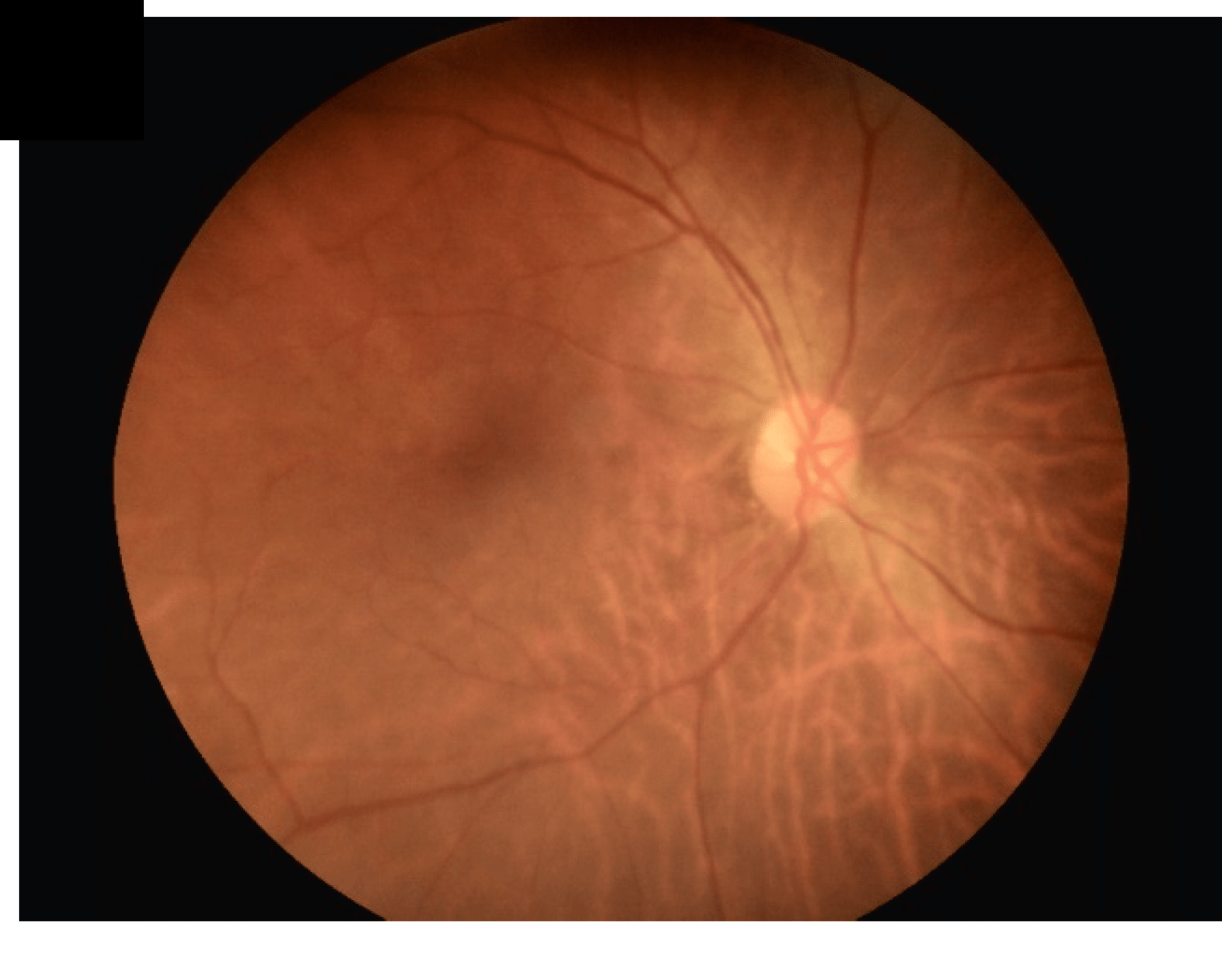
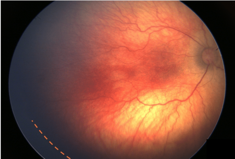
Image credits (CC BY 4.0): Left) Chan TY, Wang JH, Chen N, Chiu CJ. The Assessment of Retinal Image Quality Using a Non-Mydriatic Fundus Camera in a Teleophthalmologic Platform. Diagnostics. 2024;14(14):1543. (DOI, part of Figure 4A used) / Right) Khamees A, Schöneberger V, Kaya S, Guthoff T, Geerling G, Guthoff R. Waiting is worthwhile: ROP stage 5 with stage regression due to retinal reattachment after scleral buckling surgery (encircling band) – a case report with review of the literature. BMC Ophthalmology. 2025;25. (DOI, part of Figure 1A used) Both are the original authors’ photographs, cropped, and reproduced under theCC BY 4.0license under which they were published.
International classification (ICROP) and severity
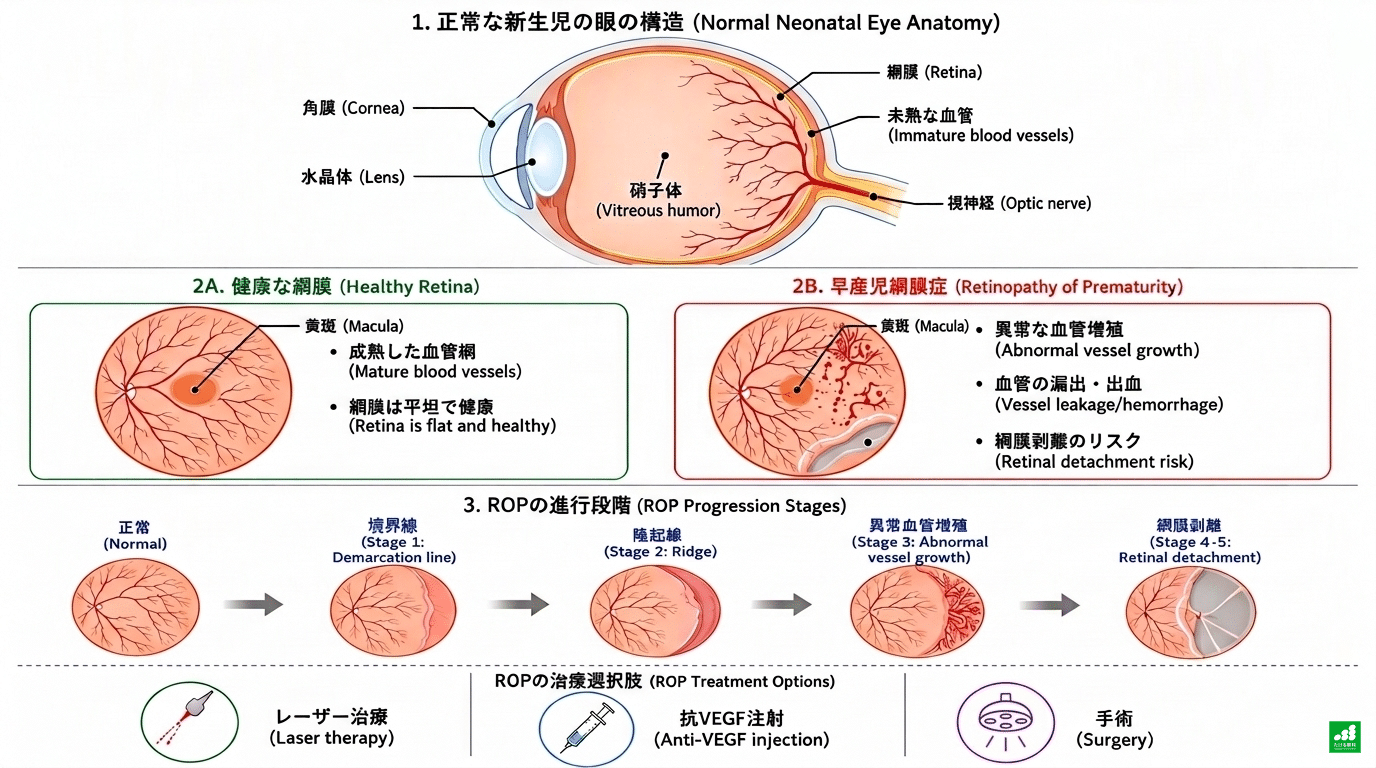
The severity of ROP is described with the International Classification of Retinopathy of Prematurity (ICROP)³. The third edition, revised in 2021, combines several criteria.
One is the zone (I–III; the closer to the optic nerve, the more posterior), which shows where the lesion lies in the retina. Posterior lesions such as Zone I require more careful assessment. The other is the stage (1–5; higher numbers mean more advanced disease, and stages 4 and 5 involve retinal detachment), which shows how far the disease has progressed.
In addition, the presence of “plus disease”—dilated, tortuous blood vessels that signal how active the disease is—is also an important factor in treatment decisions³.
📌 We use these two yardsticks, zone and stage, because future risk depends on where and how much ROP has occurred. Lesions close to the optic nerve (Zone I) are rare but can progress rapidly and call for closer follow-up.
The three axes above are summarized in the table below.
| Axis | Description |
|---|---|
| Zone (I–III) | Location of the lesion in the retina. Posterior lesions such as Zone I, close to the optic nerve, require more careful assessment |
| Stage (1–5) | Stage of disease progression. Higher numbers mean more advanced disease; stages 4 and 5 involve retinal detachment |
| Plus disease | Dilated, tortuous blood vessels and similar findings that signal how active the disease is. An important factor in treatment decisions |
In Japan, a classification developed by a research group of the former Ministry of Health and Welfare has also been used for a long time. It divides cases into slowly progressing Type I and rapidly progressing Type II (a Japan-specific classification that differs from the Type 1 and Type 2 used in international clinical trials), and describes the later course in cicatricial stages 1–5.
If your medical record or referral letter says something like “Type I” or “cicatricial stage 3,” it is based on this classification.
Screening criteria in Japan
According to the Japanese Association for Pediatric Ophthalmology,a birth weight of 1,800 g or lessora gestational age of 34 weeks or lessqualifies a baby for screening (a fundus examination in the newborn period)⁴.
The shorter the gestational age and the lower the birth weight, the higher the incidence; it is explained that about 60% of infants under 1,500 g, and nearly all infants born before 28 weeks, show some degree of ROP⁴.
Most cases resolve on their own, but some progress to a stage that needs treatment. Which course you had is often recorded in your maternal and child health handbook or in the referral letter from the neonatal unit, so it helps to check this when you see an eye doctor as an adult.
History of treatment and long-term risks that differ by generation
Treatment of ROP has changed greatly over the past several decades. Because the points to watch in adulthood differ slightly depending on the era of treatment, it is useful to know which era your treatment belonged to.
The era of cryotherapy
Cryotherapy (freezing the peripheral retina to suppress abnormal blood vessel growth) was established in the 1980s.
A multicenter study followed this generation for 15 years in severe cases that had reached threshold. It reported that treated eyes had better visual acuity and retinal structure than untreated eyes, but also that new retinal folds and retinal detachment were seen in both groups by the mid-teens⁵.
In other words, while the effect of treatment itself is clear, in severe ROP, changes in the peripheral retina and late retinal detachment can occur years later regardless of whether treatment was received.
The era of laser photocoagulation
From the 1990s, laser photocoagulation, a less burdensome treatment, became the mainstream. Through the ETROP trial that established treatment criteria⁶ and subsequent reports of visual outcomes⁷, laser treatment remains one of the widely used standard treatments today.
As for long-term outcomes in the laser generation, a study that followed 28 eyes treated with laser at threshold for 17 years reported that nearly all had myopia and that 43% (12 eyes) had progressed to high myopia (spherical equivalent worse than −6.0 diopters) at age 17⁸.
The study also showed that multiple factors, such as the shape of the cornea and lens, are involved in high myopia in these eyes, so it is not always only the axial length that is elongated, as in ordinary myopia. In any case, high myopia tends to put strain on the retina, so these eyes remain subjects of continued observation in adulthood.
There are also sobering reports on visual outcomes. In the visual outcomes of the ETROP trial, which tested the effect of early treatment of high-risk prethreshold ROP, only about one third of early-treated eyes achieved visual acuity of 0.5 (20/40) or better at age 6⁷,¹⁷.
The era of anti-VEGF treatment
From the 2010s, treatment with intraocular injection of anti-VEGF drugs (drugs that suppress abnormal blood vessel growth) spread. A trial using bevacizumab⁹ and then an international trial using ranibizumab¹⁰ reported that anti-VEGF treatment is effective in suppressing disease activity and for retinal structure.
On the other hand, it is also true that in the anti-VEGF generation, late reactivation—in which disease activity returns some time after treatment—can occur, and that lifelong long-term data have not yet accumulated as much as for laser treatment¹¹.
“Treatment is over” does not mean “follow-up is over.”This applies to everyone, whatever treatment they received in whatever era.
Of the anti-VEGF drugs, those approved in Japan for ROP are ranibizumab (Lucentis®) and aflibercept (Eylea®)¹²,¹³. Bevacizumab, which has been used in trials, is used off-label for ROP¹²,¹⁴.
If your maternal and child health handbook or referral letter records which drug was used, it is a good idea to check.
Long-term risks by treatment (table)
The table below summarizes the long-term risks for each treatment.
| Treatment | Main long-term risks and features | Source |
|---|---|---|
| Cryotherapy (from the 1980s) | At 15 years, treated eyes had better visual acuity and structure than untreated eyes. However, in severe cases, new retinal folds and late retinal detachment can appear by the mid-teens whether or not treatment was given | Ref. 5 |
| Laser photocoagulation (from the 1990s) | In a 17-year follow-up of 28 eyes treated with laser for threshold ROP, all had myopia and 43% progressed to high myopia | Ref. 8 |
| Anti-VEGF treatment (from the 2010s) | Efficacy for disease activity and retinal structure has been reported, but the risk of late reactivation and the accumulation of long-term data remain open issues. Approved drugs in Japan: ranibizumab and aflibercept | Refs. 9, 10 (efficacy); Refs. 12–14 (approval in Japan) |
What to watch for in adulthood
The following changes have been reported as more likely to occur in adulthood in people with a history of ROP. None of them is certain to happen; they are reported to occur more often than in the general population.
Myopia and high myopia
As noted above, myopia is common in people who received laser treatment at threshold, and some progress to high myopia⁸. High myopia means more than strong glasses or contact lens prescriptions: it can come with changes in the internal structure of the eye, such as the shape of the cornea and lens. Changes in the retina itself also need attention regardless of the degree of myopia, and are linked to the risk of retinal tears and retinal detachment described next.
If you notice changes such as a sudden change in prescription or extremely poor vision in one eye only, we recommend having the cause checked by an eye doctor. For a sudden change, it is important to examine not only refraction but also the state of the retina promptly.
Retinal tears and retinal detachment
Retinal tears and retinal detachment can occur in adulthood whether or not treatment was given¹,¹¹. A review of the sequelae of ROP from adolescence to adulthood also lists peripheral retinal changes and tractional and rhegmatogenous retinal detachment as long-term complications¹.
If you notice a sudden increase in floaters (spots or threads drifting in your vision), photopsia (flashes of light at the edge of your vision), or part of your visual field missing, contact an eye clinic or be seen immediately, without waiting for your next scheduled visit.
These are symptoms of possible retinal tears or retinal detachment that apply equally to people without a history of ROP.
Glaucoma
Risk of glaucoma (a disease in which the optic nerve is damaged, for example by raised eye pressure, and the visual field gradually loses areas) has also been reported¹,².
A nationwide cohort study in Taiwan of preterm infants born between 2009 and 2012 showed that people with a history of ROP had a clearly higher risk of glaucoma than those born at term (compared with preterm infants without ROP, however, no statistically clear difference was shown)². Both sets of data follow children only through childhood. The risk of glaucoma from adolescence into adulthood has been reported in a review covering people with a history of ROP in general¹.
Glaucoma often progresses with almost no symptoms, so regular eye pressure and optic nerve examinations, even when you have no symptoms, lead to early detection.
Warning signs and what to do (summary)
For the three changes above, the table below summarizes the warning signs and what to do.
| Change / complication | Warning signs | What to do |
|---|---|---|
| Myopia and high myopia | Sudden change in prescription; extremely poor vision in one eye only | Have the cause checked by an eye doctor (see one early for a sudden change) |
| Retinal tear / retinal detachment | Sudden increase in floaters, flashes of light, part of the visual field missing | See an eye doctor immediately, without waiting for your next scheduled visit |
| Glaucoma | Often almost no symptoms | Have regular eye pressure and optic nerve examinations even without symptoms |
Links with development and general health
This is somewhat specialized, but some studies have examined the relationship between a history of ROP and cognitive and motor development.
A pooled analysis of multiple studies showed a tendency for such developmental problems to be reported more often in people with ROP, but it also states clearly that the certainty of the reported evidence is very low overall¹⁵.
This does not mean that developmental problems always occur with ROP. Several factors, such as preterm birth and general health, are involved, and a causal relationship with ROP itself has not been established. For development of the whole body, not only the eyes, it is important to continue follow-up with a pediatrician.
Follow-up in childhood
Infancy and early childhood
In infancy and early childhood, visits at relatively short intervals may be needed to detect amblyopia (a condition in which the development of vision is impaired) early and to detect fast-progressing refractive errors (myopia, hyperopia, astigmatism). How many months apart is appropriate varies widely with severity and treatment history, so it is decided in consultation with your doctor.
At our clinic, for cases whose acute-phase disease has settled and who have moved on to long-term follow-up, we use a visit every few months as one guide. This is only our clinic’s guide, not a uniform standard.
Just after treatment, or while disease activity remains, much shorter intervals (several days to a week) are needed.
School age and later
From school age, for people whose condition is stable, our guide is in many cases a visit about once a year. This frequency is also not uniform; it can be shorter during periods of rapid myopia progression or while strabismus or amblyopia is being treated.
Screening guidelines jointly issued by the American Academy of Pediatrics and other organizations also recommend continued ophthalmic follow-up for children after the cicatricial stage¹⁶.
What we would like parents to know
We would like parents to know the signs of retinal detachment, such as a sudden drop in vision or floaters. Because children often cannot put their symptoms into words, please also watch for changes such as a child who suddenly seems to have trouble seeing the TV or blackboard, or who closes one eye to see.
Being able to ask for advice as soon as you notice a change is as important as regular visits. Having a regular eye doctor makes it easier to do so.
Guide to visit frequency by age
The visit guidelines above are summarized in the table below.
| Period | Visit guide (at our clinic) | Main checks |
|---|---|---|
| Just after treatment / while disease activity remains | Every few days to once a week | Checking disease activity |
| Infancy and early childhood (after disease activity has settled) | About every few months, as a guide | Early detection of amblyopia; detecting fast-progressing refractive error |
| School age and later (if stable) | About once a year, as a guide | Myopia progression, management of strabismus and amblyopia, and fundus and eye pressure checks as needed |
What we do at Takeru Eye Clinic
Even a small clinic can make good use of limited medical resources and set priorities in care. When a child with a history of ROP visits, we check the following items.
Tests are carried out by ourcertified orthoptists.
- Refraction test: highly accurate refraction testing using a cycloplegic agent (eye drops that relax the focusing muscles)
- Fundus examination: we choose between a detailed fundus examination under dilation, ornon-dilated imaging with Mirante®, depending on the child’s condition
- Evaluation of strabismus and amblyopia: we regularly check binocular vision and eye alignment
- Eye pressure check: a test that keeps glaucoma risk in mind
Of these, we give particular priority to refraction testing and fundus examination under dilation. If we judge that advanced imaging or surgical treatment is needed, we refer patients to partner institutions such as Kyushu University Hospital.

A message to patients
ROP is a disease that needs treatment or monitoring in the newborn period, and in many cases it is also a disease whose effects on the eyes need to be watched into adulthood. Even if nothing looks different on the outside, we would like you to know that changes can appear in the retina over a long period of time.
Even as an adult, we ask that you have an eye examination about once a year, depending on your treatment history and the condition of your eyes. We believe this is the simplest preparation you can make now to protect your vision for the long term.
For parents, too, continuing your child’s follow-up helps protect how they will see in the future.
References
- Özdemir HB, Özdek S. Late sequelae of retinopathy of prematurity in adolescence and adulthood. Saudi J Ophthalmol. 2022;36(3):270-277. https://pmc.ncbi.nlm.nih.gov/articles/PMC9583352/
- Huang CY, Tou SI, Lin HJ, Yen HR. Risk of long-term ophthalmological complications in Taiwanese preterm infants: a nationwide cohort study. BMJ Paediatr Open. 2024;8(1):e002279. https://pmc.ncbi.nlm.nih.gov/articles/PMC11605834/
- Chiang MF, Quinn GE, Fielder AR, et al. International Classification of Retinopathy of Prematurity, Third Edition. Ophthalmology. 2021;128(10):e51-e68. https://pubmed.ncbi.nlm.nih.gov/34247850/
- Japanese Association for Pediatric Ophthalmology. About retinopathy of prematurity (information for the general public) (in Japanese).http://www.japo-web.jp/info_ippan_page.php?id=page14
- Palmer EA, Hardy RJ, Dobson V, et al; Cryotherapy for Retinopathy of Prematurity Cooperative Group. 15-Year Outcomes Following Threshold Retinopathy of Prematurity: Final Results From the Multicenter Trial of Cryotherapy for Retinopathy of Prematurity. Arch Ophthalmol. 2005;123(3):311-318. https://pubmed.ncbi.nlm.nih.gov/15767472/
- Early Treatment for Retinopathy of Prematurity Cooperative Group. Revised Indications for the Treatment of Retinopathy of Prematurity: Results of the Early Treatment for Retinopathy of Prematurity Randomized Trial. Arch Ophthalmol. 2003;121(12):1684-1694. https://pubmed.ncbi.nlm.nih.gov/14662586/
- Good WV, Hardy RJ, Dobson V, et al; Early Treatment for Retinopathy of Prematurity Cooperative Group. Final Visual Acuity Results in the Early Treatment for Retinopathy of Prematurity Study. Arch Ophthalmol. 2010;128(6):663-671. https://pubmed.ncbi.nlm.nih.gov/20385926/
- Chou YB, Wang AG, Yang HY, Chen KJ, Yang CS. Refractive status, biometric components, and functional outcomes of patients with threshold retinopathy of prematurity: systemic review and a 17-year longitudinal study. Graefes Arch Clin Exp Ophthalmol. 2022;260(12):3809-3816. https://pubmed.ncbi.nlm.nih.gov/35729410/
- Mintz-Hittner HA, Kennedy KA, Chuang AZ; BEAT-ROP Cooperative Group. Efficacy of Intravitreal Bevacizumab for Stage 3+ Retinopathy of Prematurity. N Engl J Med. 2011;364(7):603-615. https://pubmed.ncbi.nlm.nih.gov/21323540/
- Stahl A, Lepore D, Fielder A, et al. Ranibizumab versus laser therapy for the treatment of very low birthweight infants with retinopathy of prematurity (RAINBOW): an open-label randomised controlled trial. Lancet. 2019;394(10208):1551-1559. https://pubmed.ncbi.nlm.nih.gov/31522845/
- Li JQ, Pfeil JM, Stahl A, Krohne TU. [Late sequelae of retinopathy of prematurity in infancy]. Ophthalmologie. 2023;120(6):588-596. (German-language review, English abstract) https://pubmed.ncbi.nlm.nih.gov/37221277/
- Japanese Ophthalmological Society, Committee on Ophthalmic Management of Retinopathy of Prematurity. Guide to anti-VEGF therapy for retinopathy of prematurity. Revised December 2020 (in Japanese).https://www.nichigan.or.jp/Portals/0/resources/member/guideline/rop.pdf
- Pharmaceuticals and Medical Devices Agency (PMDA). Eylea® intravitreal injection package insert (including retinopathy of prematurity). Revised April 2026 (6th edition) (in Japanese).https://www.pmda.go.jp/PmdaSearch/iyakuDetail/ResultDataSetPDF/630004_1319405A1027_1_17
- Pharmaceuticals and Medical Devices Agency (PMDA). Lucentis® intravitreal injection review report (indication added: retinopathy of prematurity). November 2019 (in Japanese).https://www.pmda.go.jp/drugs/2019/P20191115001/300242000_22600AMX00565_A100_1.pdf
- Diggikar S, et al. Retinopathy of prematurity and neurodevelopmental outcomes in preterm infants: a systematic review and meta-analysis. Front Pediatr. 2023;11:1055813. https://pubmed.ncbi.nlm.nih.gov/37009271/
- Fierson WM; American Academy of Pediatrics Section on Ophthalmology, American Academy of Ophthalmology, American Association for Pediatric Ophthalmology and Strabismus, American Association of Certified Orthoptists. Screening Examination of Premature Infants for Retinopathy of Prematurity. Pediatrics. 2018;142(6):e20183061. https://pubmed.ncbi.nlm.nih.gov/30478242/
- Hong EH, Shin YU, Cho H. Retinopathy of prematurity: a review of epidemiology and current treatment strategies. Clin Exp Pediatr. 2022;65(3):115-126. https://doi.org/10.3345/cep.2021.00773