![Takeru Eye Clinic | Takatori Shopping Street, Sawara-ku, Fukuoka City [Nishijin Station / Fujisaki Station]](https://takeru-eye.com/wp-content/uploads/2022/10/takeru_logo_for-WP-header.png)

When the clinic opened in October 2018, we introduced OCT angiography (OCTA).
Because I had been involved in research on retinal disease, uveitis, and glaucoma, I kept negotiating to bring in OCTA from the autumn of 2017, while the clinic was still being prepared.
OCTA was newly added to the list of insured examinations in April 2018, about six months before we opened.
We started with the RS-3000 Advance2 (NIDEK) and later upgraded to the Mirante, which can also take ultra-widefield fundus images.

An examination that shows blood flow inside the eye without a contrast dye
OCTA turns the flow in the fine blood vessels at the back of the eye into an image without injecting a contrast dye into an arm vein. One scan takes only a few seconds.
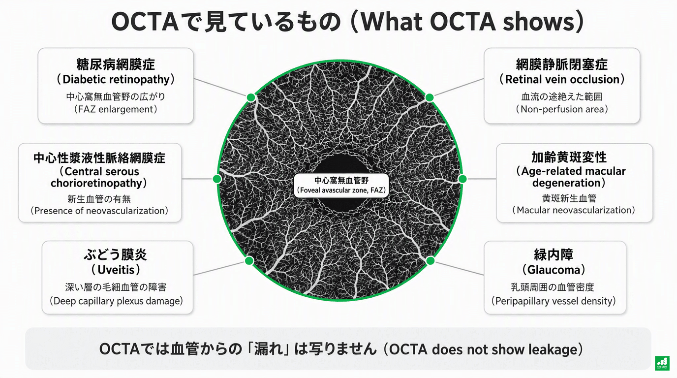
What we look at depends on the disease.
In diabetic retinopathy, we check where the blood vessels break off in the center of vision. In retinal vein occlusion, we check the blood flow beyond the blocked vessel.
In age-related macular degeneration, we look for vessels that should not be there. In glaucoma, we measure the density of vessels around the optic nerve.
In uveitis, we look at the effects that inflammation has left on the vessels.
On the other hand, OCTA does not show “leakage” from vessels. When we need to know how active inflammation or new vessels are right now, a dye-based test may still be necessary.
Until now, dye-based tests were the standard
Until now, to examine the fine vessels at the back of the eye, the main method was to inject a contrast dye into an arm vein and photograph the eye with a fundus camera (fluorescein angiography).
The dye can cause side effects such as nausea and hives, and rarely shock.
The test also takes time before and after, and an appointment had to be made in advance.
OCTA scans the same spot repeatedly and extracts only the changes in signal caused by red blood cells moving inside the vessels.1Because no contrast dye is used, the dye injection and its side effects are avoided.
One scan takes only a few seconds. At our clinic you do not need to book a separate day for OCTA; it can be done on the spot during your visit.
However, we sometimes need to repeat a scan depending on eye movement or image quality, and depending on conditions we may use eye drops to dilate the pupil.
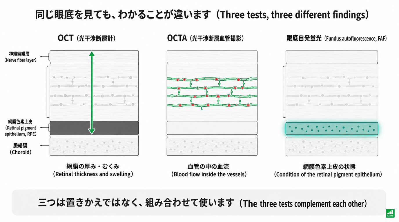
OCT, OCTA, and fundus autofluorescence: the names are similar, but they look at different things
“OCT” examines a cross-section of the retina to measure thickness and swelling. OCTA adds blood flow information to it.
They use the same device and you sit the same way, but the images mean different things.
There is also a test called fundus autofluorescence (FAF). It checks whether waste products are accumulating in the retinal pigment epithelium, the outermost cell layer of the retina, and whether cells have been lost.2。
OCTA, which shows blood flow, and FAF, which shows the condition of cells, are not interchangeable. Each has its own role depending on the disease.
The key point is that OCTA cannot directly show “leakage” from blood vessels.
Seeing a vessel is not the same as knowing that fluid is leaking from it now. We decide by combining OCT, the examination, and, when needed, a dye-based test.1。
Diabetic retinopathy: examining the vessels at the center of vision
In diabetic retinopathy, the retinal capillaries can be damaged so that blood reaches the retina less easily.
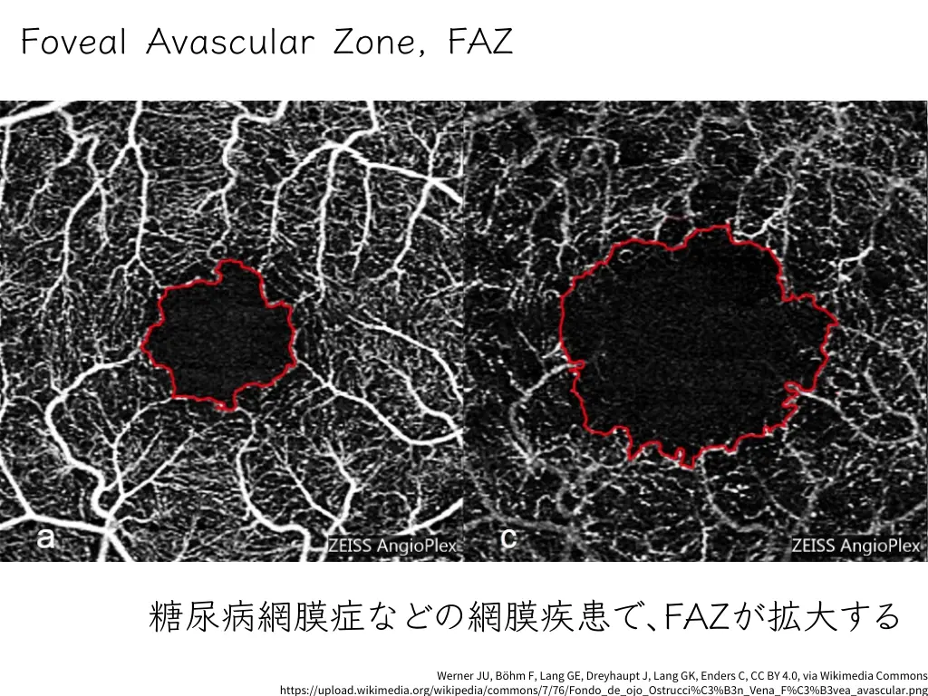
One thing we often evaluate at our clinic is the foveal avascular zone (FAZ). At the center of vision there is a small area that normally has no capillaries.
We examine its size and shape and where the surrounding vessels break off.
Checking whether the FAZ has enlarged and whether the surrounding vessels have become sparse gives clues to impaired blood flow in the part of the retina that matters for vision.3。
However, FAZ size varies from person to person, and the severity cannot be decided from a number alone.
Swelling is checked with OCT, and bleeding and the peripheral retina are checked with a fundus examination.Even when OCTA looks normal, regular fundus examinations for diabetes are still necessary.

Retinal vein occlusion: checking the blood flow beyond the blockage
Retinal vein occlusion is a disease in which a retinal vein is blocked and blood beyond the blockage cannot flow properly.
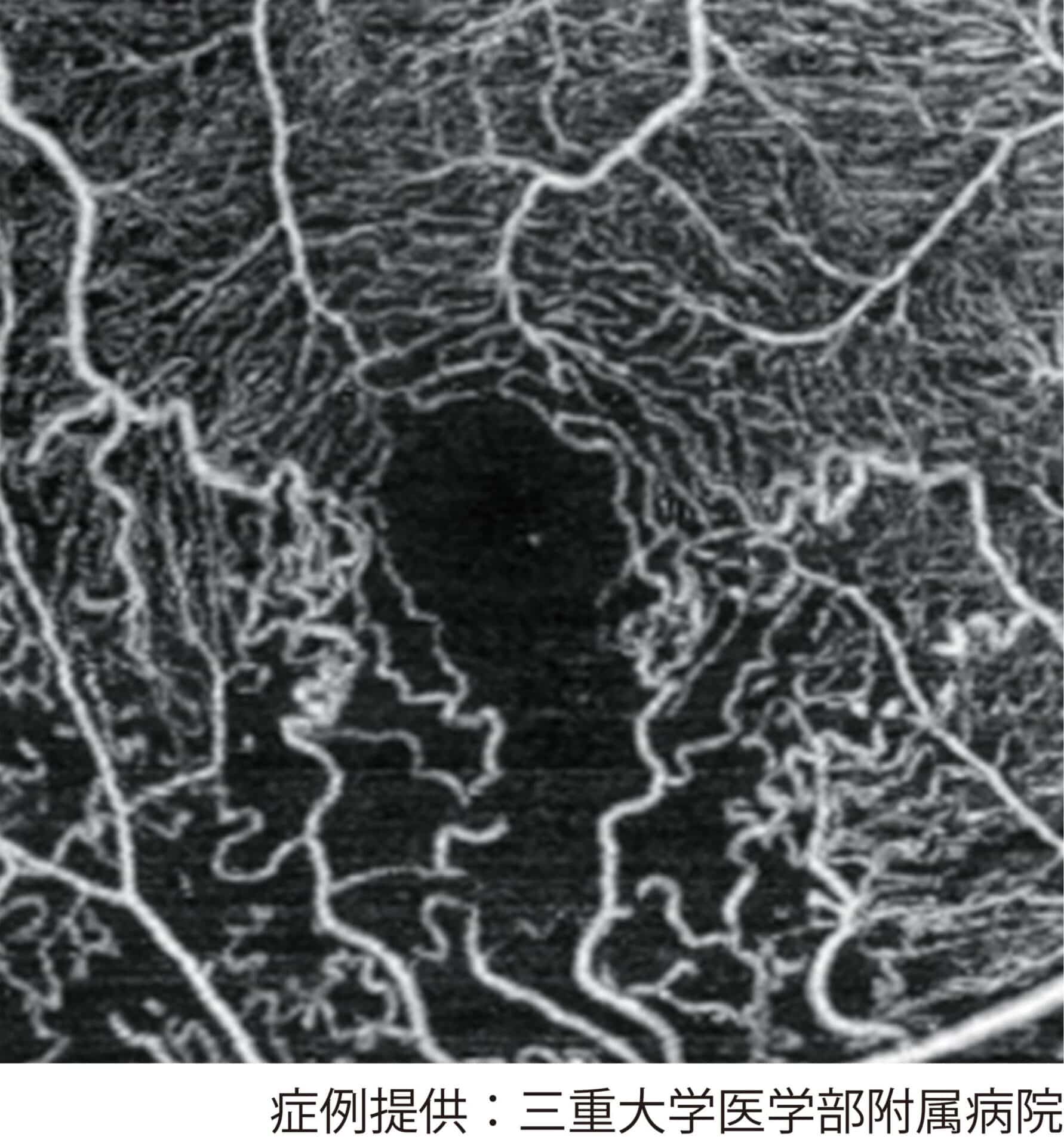
With OCTA, we can see areas where the blood flow signal is reduced and where vessels are interrupted, in both the superficial and deep layers of the retina.4。
Areas that blood does not reach, and changes in the vessels, are clues to the state of the disease4. Treatment intervals are decided together with the swelling seen on OCT, visual acuity, and the response to previous treatment.
However, a study comparing widefield OCTA with ultra-widefield angiography reported that the larger the area without blood flow, the more OCTA tends to underestimate its proportion.5。
When we need to confirm how far the lesion extends into the peripheral retina, ultra-widefield dye-based angiography may be necessary.
Age-related macular degeneration: looking for vessels that should not be there
In age-related macular degeneration, abnormal vessels can form in the macula, the center of vision. These are called macular neovascularization (MNV).
OCTA can sometimes capture the signal of blood flowing inside this new vessel without a contrast dye.
A pooled analysis of multiple studies reported that the sensitivity for detecting new vessels is about 0.83 to 0.89. The breakdown was 0.89 in an analysis of age-related macular degeneration6 and 0.83 in a separate analysis of active choroidal neovascularization.7。
Sensitivity is the proportion of eyes with new vessels that the test correctly identifies as having them.
On the other hand, the proportion of eyes without new vessels that the test correctly identifies as having none (specificity) was reported to be 0.89 to 0.966,7。
Even when a new vessel is seen, we check that it is not a false finding caused by image artifacts.
Conversely, a vessel that is not seen does not prove that there is no neovascularization. In either case, we decide together with the examination and OCT findings.
These figures come from studies with different populations and imaging conditions. They do not guarantee our clinic’s detection rate or the diagnosis for any individual.
New vessels that are not yet leaking fluid are sometimes found as well.
This can help us watch for future changes.
However, finding a vessel does not mean we start injection treatment right away.. We decide based on whether fluid is present on OCT, bleeding, visual acuity, and symptoms.8。
Glaucoma: examining the vessels around the optic nerve
Glaucoma is a disease in which the optic nerve, which carries information from the eye to the brain, is damaged and the visible field narrows.
With OCTA, we examine the optic disc, where the optic nerve leaves the eyeball, and the fine vessels around it.
“Peripapillary vessel density” is an index of what proportion of the imaged area is occupied by vessels with blood flow.
A report pooling 43 studies found that the index for distinguishing glaucomatous eyes from healthy eyes (AUROC) averaged 0.80 around the optic disc. In the regions examining the superficial vessels around the disc, divided into inferior and superior sides, the averages were 0.86 and 0.87.9。
The closer the AUROC is to 1, the better the ability to discriminate, but it does not mean that 80–87% of patients can be diagnosed correctly.
We evaluate it together with eye pressure, visual field tests, and the nerve thickness seen on OCT.
As glaucoma progresses, the nerve becomes thin, and OCT thickness values alone may make it hard to capture further change.
Even at this stage, OCTA vessel density has been reported to be a possible source of additional information.10。
On the other hand, the values also vary with the degree of myopia and image clarity. OCTA is a supplementary test and is not performed uniformly for every patient with glaucoma.

Uveitis: seeing how inflammation has affected the vessels
OCTA is also useful in my own specialty of uveitis and ocular immunology.
Uveitis is a general term for diseases in which inflammation occurs inside the eye. When inflammation reaches the retina or choroid, it can affect the fine vessels and the function of the center of vision.11。
- Behçet disease: We check whether the retinal capillaries are damaged and whether there are areas with little blood flow signal. In active Behçet disease, a report described changes in the fine vessels around the fovea more precisely than with dye-based angiography, with stronger changes in the deep layer.12。
- Vogt-Koyanagi-Harada disease (VKH): In the fine vessels of the choroid, the layer outside the retina, we can sometimes see areas where the flow signal is weak. These areas shrink with treatment, and a case has been reported in which they reappeared before a relapse after treatment was stopped.13。
- APMPPE and serpiginous choroiditis, among others: In diseases that cause white lesions in the fundus, the depth at which blood flow changes occur serves as a clue for telling the diseases apart.11。
- New vessels associated with inflammation: We check whether an abnormal vessel has developed in an inflammatory lesion.11。
OCTA alone cannot determine how intense inflammation is or whether treatment is needed.
In retinal vasculitis in particular, fluorescein angiography to check for dye leakage may be necessary.
Information is also available in other diseases
Central serous chorioretinopathy
Central serous chorioretinopathy is a disease in which fluid collects under the retina at the center of vision. Most cases resolve on their own, but when it persists, new vessels may develop.
OCTA is well suited to finding these new vessels. A pooled analysis of studies reported a sensitivity of 92.9% and a specificity of 99.4%.14。
However, in studies that used a combined judgment from multiple tests as the reference standard, the sensitivity was 78–81% and the specificity 96–97%, and the numbers also differed slightly depending on who made the judgment.15。
When treatment is being considered for a persistent case, this can reduce the number of dye-based tests in some situations.

Retinitis pigmentosa
Retinitis pigmentosa is a disease in which the cells that sense light are gradually lost.
In a study of 73 cases reported from Kyushu University, the density of blood flow around the center of vision was related to visual acuity and to results of central visual field tests.16。
Blood flow itself is not a target of treatment, but it is one of the materials for thinking about changes in vision.
In retinitis pigmentosa, fundus autofluorescence (FAF) is also useful for checking the condition and changes of the retina.2. We follow the course by combining it with visual acuity, visual field tests, OCT, and other tests, and OCTA adds information about blood flow.16。
What OCTA cannot tell us
Not every place where the blood flow signal is missing is a place where a vessel is blocked.
Vessels are also hard to image when the flow is slow, when bleeding or clouding inside the eye keeps light from reaching them, or when the eye moves.
The area that can be imaged is also limited. When swelling of the retina shifts the boundaries between layers, the appearance of the image changes as well.
Values change with different devices and analysis methods, so worsening or improvement of a disease cannot be judged only from values before and after a device is replaced.1,17。
OCTA alone cannot determine how active new vessels or inflammation are.
When needed, we ask partner medical institutions such as Kyushu University Hospital to perform a dye-based test.
Having the test and reading the results
OCTA is an insured examination when it is judged medically necessary (there are limits on how many times it can be billed).
One scan takes only a few seconds and is painless. Depending on conditions, we may use eye drops to dilate the pupil.
At our clinic, we choose the tests that are needed according to your symptoms and examination findings.
We show the results on a monitor set at the patient’s eye level while explaining them.
Where are the changes? How do they compare with last time? What should we check next? We explain the plan going forward while connecting the images to how you see.
If anything is unclear, please do not hesitate to ask.
Main references
- Spaide RF, Fujimoto JG, Waheed NK, Sadda SR, Staurenghi G. Optical coherence tomography angiography. Prog Retin Eye Res. 2018;64:1-55. https://doi.org/10.1016/j.preteyeres.2017.11.003
- Schmitz-Valckenberg S, Pfau M, Fleckenstein M, et al. Fundus autofluorescence imaging. Prog Retin Eye Res. 2021;81:100893. https://doi.org/10.1016/j.preteyeres.2020.100893
- Waheed NK, Rosen RB, Jia Y, et al. Optical coherence tomography angiography in diabetic retinopathy. Prog Retin Eye Res. 2023;97:101206. https://doi.org/10.1016/j.preteyeres.2023.101206
- Tsai G, Banaee T, Conti FF, Singh RP. Optical coherence tomography angiography in eyes with retinal vein occlusion. J Ophthalmic Vis Res. 2018;13(3):315-332. https://doi.org/10.4103/jovr.jovr_264_17
- Hafner M, Herold TR, Deiters V, von Livonius B, Priglinger SG, Gerhardt MJ. Quantitative comparison of a novel wide-field OCT-angiography device with ultrawide-field fluorescein angiography in detecting retinal nonperfusion in vascular retinopathies. BMC Ophthalmol. 2025;25(1):642. https://doi.org/10.1186/s12886-025-04468-z
- Chen CX, Liu ML, Cao K, Yusufu M, Wang JD. Diagnostic value of optical coherence tomography angiography for choroidal neovascularization in age-related macular degeneration: a systematic review and meta-analysis. Ophthalmic Res. 2021;64(5):704-712. https://doi.org/10.1159/000511265
- Wang M, Gao S, Zhang Y, Zhang M. Sensitivity and specificity of optical coherence tomography angiography in the diagnosis of active choroidal neovascularization: a systematic review and meta-analysis. Graefes Arch Clin Exp Ophthalmol. 2021;259(12):3529-3536. https://doi.org/10.1007/s00417-021-05239-4
- Yang J, Zhang Q, Motulsky EH, et al. Two-year risk of exudation in eyes with nonexudative age-related macular degeneration and subclinical neovascularization detected with swept source optical coherence tomography angiography. Am J Ophthalmol. 2019;208:1-11. https://doi.org/10.1016/j.ajo.2019.06.017
- Bekkers A, Borren N, Ederveen V, et al. Microvascular damage assessed by optical coherence tomography angiography for glaucoma diagnosis: a systematic review of the most discriminative regions. Acta Ophthalmol. 2020;98(6):537-558. https://doi.org/10.1111/aos.14392
- Rao HL, Pradhan ZS, Suh MH, Moghimi S, Mansouri K, Weinreb RN. Optical coherence tomography angiography in glaucoma. J Glaucoma. 2020;29(4):312-321. https://doi.org/10.1097/IJG.0000000000001463
- Pichi F, Sarraf D, Arepalli S, et al. The application of optical coherence tomography angiography in uveitis and inflammatory eye diseases. Prog Retin Eye Res. 2017;59:178-201. https://doi.org/10.1016/j.preteyeres.2017.04.005
- Khairallah M, Abroug N, Khochtali S, et al. Optical coherence tomography angiography in patients with Behçet uveitis. Retina. 2017;37(9):1678-1691. https://doi.org/10.1097/IAE.0000000000001418
- Aggarwal K, Agarwal A, Mahajan S, et al; OCTA Study Group. The role of optical coherence tomography angiography in the diagnosis and management of acute Vogt-Koyanagi-Harada disease. Ocul Immunol Inflamm. 2018;26(1):142-153. https://doi.org/10.1080/09273948.2016.1195001
- Kiilgaard HC, Nissen AHK, Balaratnasingam C, et al. Diagnostic accuracy of OCT angiography for macular neovascularization in central serous chorioretinopathy: a systematic review and meta-analysis. Acta Ophthalmol. 2024;102(7):749-758. https://doi.org/10.1111/aos.16739
- Ng DS, Ho M, Chen LJ, et al. Optical coherence tomography angiography compared with multimodal imaging for diagnosing neovascular central serous chorioretinopathy. Am J Ophthalmol. 2021;232:70-82. https://doi.org/10.1016/j.ajo.2021.05.029
- Koyanagi Y, Murakami Y, Funatsu J, et al. Optical coherence tomography angiography of the macular microvasculature changes in retinitis pigmentosa. Acta Ophthalmol. 2018;96(1):e59-e67. https://doi.org/10.1111/aos.13475
- Mihailovic N, Brand C, Lahme L, et al. Repeatability, reproducibility and agreement of foveal avascular zone measurements using three different optical coherence tomography angiography devices. PLoS One. 2018;13(10):e0206045. https://doi.org/10.1371/journal.pone.0206045
Revision history
- First published: October 29, 2018
- Last updated: September 22, 2026 (added disease-specific content and updated the references)