![Takeru Eye Clinic | Takatori Shopping Street, Sawara-ku, Fukuoka City [Nishijin Station / Fujisaki Station]](https://takeru-eye.com/wp-content/uploads/2022/10/takeru_logo_for-WP-header.png)

The sclera, the white part of the eye that forms the wall of the eyeball, is rich in collagen and connective tissue.
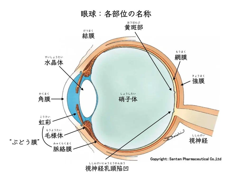
For this reason, inflammation involving collagen-containing tissue:
rheumatoid arthritis, ankylosing spondylitis, lupus
may be reflected in the eye as collagen disease:
When scleral inflammation is suspected, we also ask about
back pain, joint pain, and digestive symptoms
and similar signs.
Inflammation somewhere in the body is sometimes reflected in the eyes.
Eye Pain Caused by Scleritis
✔ Eye pain
✔ Eye discomfort
✔ Redness of the white of the eye
✔ Blurred vision
✔ Tearing
✔ Sensitivity to light
Symptoms such as eye pain and redness are sometimes caused by scleritis.
The pain of scleritis can be very severe:
It tends to worsen at night and can spread from the face to the jaw.
Why does scleritis cause eye pain?
The sclera is supplied by nerves from the cranial nerve → ciliary ganglion pathway,1and the pain is thought to arise along with the inflammation.
・Anterior scleritis (inflammation at the front of the eyeball)
・Posterior scleritis (inflammation at the back of the eyeball)
Anterior scleritis is more common and is often seen in everyday practice.
Posterior scleritis may also be complicated by
・Retinal detachment2(affecting vision)
・Angle-closure glaucoma3(raising eye pressure).
These complications can occur.
Inflammation may also involve both the anterior and posterior sclera.
Nodular (lump-like) posterior scleritis is sometimes mistaken for choroidal malignant melanoma (an eye cancer),4。
and although very rare, it can be difficult to tell them apart.
Causes of scleritis
Scleritis is often associated with systemic or autoimmune disease.
○ Arthritis (joint swelling, stiffness, pain, etc.)5
○ Lupus (systemic lupus erythematosus, SLE)6
○ Inflammatory bowel disease: ulcerative colitis and Crohn’s disease 7
○ Sjögren’s syndrome 8
○ Ankylosing spondylitis 9
○ Granulomatosis with polyangiitis 10
○ Scleroderma 11 Other collagen diseases / connective tissue diseases
○ After an eye injury: fungal or parasitic scleritis 12 and others
Scleritis care
We examine the eye with a slit-lamp microscope, OCT, and enhanced-depth imaging OCT (EDI-OCT).
We ask about your general health, and may manage your care together with a rheumatology/collagen disease department.
Treatment ranges from steroid eye drops (Rinderon®®, etc.) to cases that require surgery.
It may recur repeatedly.
After severe inflammation, the sclera can become thin (scleral thinning).
When it becomes very thin, the underlying uvea may show through:
In severe cases, the uvea can bulge out.
If that happens, serious visual impairment may remain.
1. May, C.A., 2004. Morphology of the long and short uveal nerves in the human eye. J Anat 205, 113–120. https://doi.org/10.1111/j.0021-8782.2004.00317.x
2. Kellar, J.Z., Taylor, B.T., 2015. Posterior Scleritis with Inflammatory Retinal Detachment. West J Emerg Med 16, 1175–1176. https://doi.org/10.5811/westjem.2015.8.28349
3. Heinz, C., Bograd, N., Koch, J., Heiligenhaus, A., 2013. Ocular hypertension and glaucoma incidence in patients with scleritis. Graefes Arch Clin Exp Ophthalmol 251, 139–142. https://doi.org/10.1007/s00417-012-2108-3
4. Alsharif, H.M., Al-Dahmash, S.A., 2018. Atypical posterior scleritis mimicking choroidal melanoma. Saudi Med J 39, 514–518. https://doi.org/10.15537/smj.2018.5.22130
5. Zlatanović, G., Veselinović, D., Cekić, S., Živković, M., Đorđević-Jocić, J., Zlatanović, M., 2010. Ocular manifestation of rheumatoid arthritis-different forms and frequency. Bosn J Basic Med Sci 10, 323–327.
6. Sivaraj, R.R., Durrani, O.M., Denniston, A.K., Murray, P.I., Gordon, C., 2007. Ocular manifestations of systemic lupus erythematosus. Rheumatology (Oxford) 46, 1757–1762. https://doi.org/10.1093/rheumatology/kem173
7. Troncoso, L.L., Biancardi, A.L., de Moraes Jr, H.V., Zaltman, C., 2017. Ophthalmic manifestations in patients with inflammatory bowel disease: A review. World J Gastroenterol 23, 5836–5848. https://doi.org/10.3748/wjg.v23.i32.5836
8. Ahmadi-Simab, K., Lamprecht, P., Nolle, B., Ai, M., Gross, W., 2005. Successful treatment of refractory anterior scleritis in primary Sjögren’s syndrome with rituximab. Ann Rheum Dis 64, 1087–1088. https://doi.org/10.1136/ard.2004.027128
9. Bacchiega, A.B.S., Balbi, G.G.M., Ochtrop, M.L.G., de Andrade, F.A., Levy, R.A., Baraliakos, X., 2017. Ocular involvement in patients with spondyloarthritis. Rheumatology (Oxford) 56, 2060–2067. https://doi.org/10.1093/rheumatology/kex057
10. Sfiniadaki, E., Tsiara, I., Theodossiadis, P., Chatziralli, I., 2019. Ocular Manifestations of Granulomatosis with Polyangiitis: A Review of the Literature. Ophthalmol Ther 8, 227–234. https://doi.org/10.1007/s40123-019-0176-8
11. de A. F. Gomes, B., Santhiago, M.R., Magalhães, P., Kara-Junior, N., de Azevedo, M.N.L., Moraes, H.V., 2011. Ocular findings in patients with systemic sclerosis. Clinics (Sao Paulo) 66, 379–385. https://doi.org/10.1590/S1807-59322011000300003
12. Guerrero-Wooley, R.L., Peacock, J.E., 2018. Infectious Scleritis: What the ID Clinician Should Know. Open Forum Infect Dis 5. https://doi.org/10.1093/ofid/ofy140